

-
Termékek

-
Automatizált és klasszikus mikroszkópia
-
Automatizált mikroszkópia
-
ImageXpress automatizált mikroszkópia
- ImageXpress Micro Confocal High-Content Imaging System
|
|
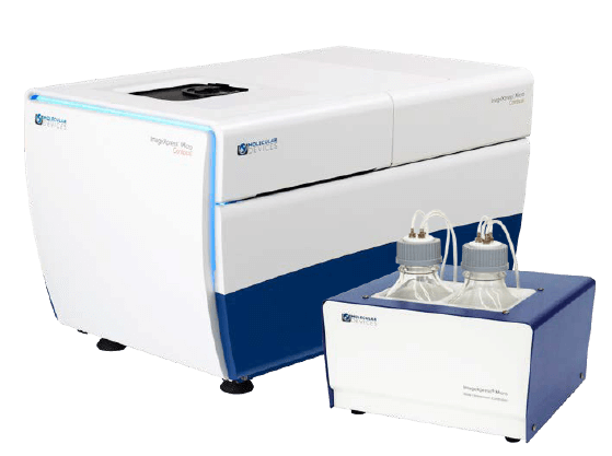
ImageXpress Micro Confocal High-Content Imaging System |


The ImageXpress® Confocal system is a high-content solution that can switch between widefield and confocal imaging of fixed and live cells. It can capture high quality images of whole organisms, thick tissues, 2D and 3D models, and cellular or intracellular events. The spinning disc confocal and sCMOS camera enable imaging of fast and rare events like cardiac cell beating and stem cell differentiation. With the MetaXpress software, the system enables many confocal imaging applications from 3D assay development to screening.
ÁRAJÁNLATOT, INFORMÁCIÓT KÉREK!
High-content imaging solution with proprietary spinning disc confocal
The ImageXpress Micro Confocal High-Content Imaging System lets you capture research-quality images with the widest range of objective lenses, allowing you to work at the resolution appropriate for your biology, including whole organism, thick tissues, 3D spheroid assays, and cellular or intracellular events—at a speed you would only expect from widefield screening.
The ImageXpress Micro Confocal system combines the ability to capture high-quality, publication-grade images without sacrificing throughput, reliability, or flexibility. Quickly and easily switch between confocal and widefield imaging modes to satisfy the specific throughput and sensitivity needs for your assay. Additionally, you can choose from a range of available confocal disk geometries to select the best fit for your experiments.
Combined with MetaXpress® High-Content Image Acquisition and Analysis Software, the ImageXpress Micro Confocal system provides you with a complete multi-dimensional, high-throughput screening solution to help you discover your next landmark scientific breakthrough.
For researchers looking to expand their laboratory’s capabilities, the ImageXpress Micro Confocal system leverages large field-of-view optics to map macro structures with minimal tiling. In addition, querying of large cell populations is accelerated, speeding up the characterization of highly heterogeneous samples or identification of rare sub-populations.
Built on over 30 years of cell-based imaging experience, the ImageXpress Micro Confocal High-Content Imaging system is engineered for performance to reliably accelerate the pace of your research.
|
|
Match acquisition mode to your assay needs with multiple confocal geometries and widefield mode. Quickly and easily switch between widefield and several confocal disk configurations, including an exclusive slit option, to capture high quality images at high-throughput speeds. |
|
|
Capture publication-quality images without sacrificing throughput using our proprietary AgileOptix™ technology. Our proprietary AgileOptix™ technology provides increased sensitivity resulting in high contrast and high uniformity of intensity and shape across a large field |
|
|
Acquire statistically relevant data faster with large field of view and >3 log dynamic range. Featuring a high-quantum efficiency sCMOS detector and power-optimized solid-state light source, the ImageXpress Micro Confocal system delivers the images you need at the throughput of widefield systems. |
|
|
Expand your research capabilities with available options. Available options include Environmental Control (temperature, humidity and CO2 control for multi-day live-cell experiments), Transmitted Light True Phase contrast option and Fluidics (on board disposable pipetting). |


|
Investigate every aspect of a cellular pathway
Combined with MetaXpress High-Content Image Acquisition and Analysis Software, the ImageXpress Micro Confocal system is a complete solution that enables you to interpret your images, understand your data, and explore new ideas—in both widefield and confocal modes. |
|
|
Automatically acquire images 1-100x objective magnification in slides and multi-well plates up to 1536-well format. Acquire >160,000 wells per day at 4x magnification with the system’s unique multi-well crop feature. A 10X objective allows the entire well of a 1536-well plate to be imaged in one field-of-view while increasing magnifications allows the resolution of fine detail for analysis of subcellular events. High resolution and dynamic range
Our proprietary AgileOptix™ technology, provides increased sensitivity resulting in high contrast and high uniformity of intensity and shape across a large field of view. This optical path also achieves near-theoretical Z resolution. Featuring a high-quantum efficiency sCMOS detector and power-optimized solid-state light source, the ImageXpress Micro Confocal system delivers the images you need at the throughput of widefield systems. Quickly and easily switch between confocal and widefield imaging modes to satisfy the specific throughput and sensitivity needs for your assay. Additionally, you can choose from a range of available confocal disk geometries to select the best fit for your experiments, including an exclusive slit option to capture high quality images at high-throughput speeds. |
|


Műszaki specifikációk
General specifications
>4 megaPixel sCMOS Camera
1.96 mm2 Field of View with 10x objective
>200,000 wells/day
Air (1X-100X), Oil (1X-100X), and Water (20X, 40X, and 60X)
High intensity laser option for brighter signal
8 position emission wheel and 5 position dichroic filter wheel
Laser + image autofocus
Phase Contrast
Transmitted Light
Brightfield
Confocal and Widefield mode
Plates supported Up to 1536-well plates
Microscope slides
Environmental Control: Temperature, CO2 and humidity
Liquid handling
Robotics/Automation
MetaXpress Software with Custom Module Editor
MD Store Data Management
Applikációk
Applications of ImageXpress Micro Confocal High-Content Imaging System
-

Organ-on-a-Chip Assays
Emulating organ physiology by co-culturing cells in a supportive 3D matrix and using microfluidic channels to perfuse nutrients or compounds over the resulting cellular structures, is rapidly gaining popularity as a biologically relevant screening model for new drugs or toxicity. The ImageXpress system provides a turnkey solution for both 2D or 3D applications and can accommodate many different types of slide and plate formats.
-

Neurite Outgrowth / Neurite Tracing
Neurons create connections via extensions of their cellular body called processes. This biological phenomenon is referred to as neurite outgrowth. Understanding the signaling mechanisms driving neurite outgrowth provides valuable insight into neurotoxic responses, compound screening, and for interpreting factors influencing neural regeneration. Using the ImageXpress Micro system in combination with MetaXpress Image Analysis Software automated neurite outgrowth imaging and analysis is possible for slide or microplate-based cellular assays.
-

Spheroid Assays
Three-dimensional (3D) cells cultured into spheroids provide a more complete and physiologically predictive model especially for cancer research. Acquiring measurements from the larger structure involves acquiring images from different depths within the body of the spheroid and analyzing them in 3D or collapsing into a single 2D stack before analysis. The ImageXpress Micro system and MetaXpress software are designed to make acquisition and analysis of 3D spheroids fast and accurate.
-

3D Cell Imaging and Analysis
Three-dimensional (3D) cell models are physiologically relevant and more closely represent tissue microenvironments, cell-to-cell interactions, and biological processes that occur in vivo. Now you can generate more predictive data by incorporating technologies like the ImageXpress system with the integrated 3D Analysis Module in MetaXpress software. This single interface will enable you to meet 3D acquisition and analysis challenges without compromise to throughput or data quality, giving you confidence in your discoveries.
-

3D Gel Matrix Assays
The development and integration of three-dimensional (3D) assay models are becoming popular to drive translational biology. Our ImageXpress system allows superior visualization of thick samples by minimizing background fluorescence and increasing sharpness, resulting in accurate image segmentation. Increase the biological relevance of your screening assays with a seamless integration between acquisition and analysis of cells in a 3D space to yield volume, intensity, and distance measurements.
-

Toxicity Screening
Screening for off-target or toxic effects is very important during the development of new drugs and for the extension of the therapeutic potential of existing molecules. ImageXpress systems are fully integrated hardware and software platforms for automated acquisition and analysis of images for high-throughput cell-based cytotoxicity testing. Configured with optional environmental control, living cell responses or kinetic reactions can be monitored in real time for several days.
Videók, ismertetők
![]() Forgalmazott termékeink gyártói - keressen gyártó szerint a logóra kattintva
Forgalmazott termékeink gyártói - keressen gyártó szerint a logóra kattintva
